Discover the Anatomy of the Eye
Iris
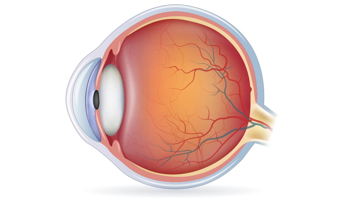
The iris is the colored, ring-shaped membrane surrounding the pupil. The connective tissues and smooth muscle fibers adjust themselves to allow light into the eye by shrinking or widening the pupil. When people ask what color eyes you have, they are asking what color your irises are!
Cornea
The cornea is the clear structure at the front of the eye that makes up the central, outer surface. It is like the windshield of a car allowing light inside and protecting the iris and pupil. And just like a windshield, if it becomes cloudy or opaque, it can significantly limit a person’s ability to see.
Pupil
The pupil is a dark circular opening in the center of the iris. In conjunction with the iris, the pupil allows light to enter the eye so that it can be focused on the retina and begin the process of sight. When the iris works to make the pupil larger in low-light situations, it is allowing as much light in as possible so that you can have better night vision. On the other hand, when the iris works to make the pupil smaller in high-light situations, it is restricting the amount of light in to avoid a glare, discomfort, or even damage.
Lens
The lens is an elastic, transparent tissue behind the iris. It changes shape to bend and focus light, forming a clear image in the back of the eye. It expands to focus on close objects and becomes thinner to focus on distant objects.
Retina
The retina is the light-sensitive membrane that lines the inner surface of the back of the eye. It is made up of layers of cells, including a layer of millions of photoreceptor cells. They gather the light that is focused by the cornea and lens and use it to produce images. This combination of nervous and chemical signals are transported through the optic nerve to the visual centers of the brain for interpretation. The signals are then converted into images of what you visually see every day!
Optic Nerve
The optic nerve is a cable-like arrangement of nerve fibers at the back of the eye that connects your brain to the eye. By receiving light signals from about 125 million photoreceptor cells in the retina, the optic nerve transfers the resulting impulses to the brain and then interprets them as images.
Vitreous humor
The vitreous humor is the colorless, jelly-like substance that fills the space inside the eye from the lens to the retina. It is made up of 99% water as well as collagen, protein, salts, and sugars. It helps maintain the shape of the eyeball and protects the retina from sliding out of place.
Sclera
The sclera, commonly known as the “white part” of the eye, is made up of dense, fibrous connective tissue that extends over more than 80% of the eye’s surface. Its main function is to help protect the eye. For example, the tough tissues shield the eye from external trauma and help it maintain its shape. The sclera also assists in the movement of the eye by providing a sturdy attachment for the extraocular muscles.
Ciliary body
The ciliary body is the circular band of muscles that sits behind the iris. These muscles change the shape of the lens in order to modify the eye’s focus. The ciliary body also secretes the aqueous fluid that fills the chambers and provides nutrition to the tissues in the eyes.
Choroid
The choroid is the thin layer of tissue that lies between the retina (the inner layer of nerve tissue toward the back of the eye) and the sclera (the outer layer that makes up the white of the eye). It is filled with blood vessels that carry oxygen and nourishment to the adjacent outer retina as well as to the rest of the eye.
At SureVision Eye Centers,
Vision Like You’ve Never Seen Before™
The surgeons and doctors at SureVision Eye Centers are among the most experienced and well-trained in the world. Each year, they perform thousands of surgical and vision correction procedures. At SureVision Eye Centers, we believe everyone should have a lifetime of their best possible vision.


